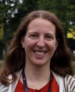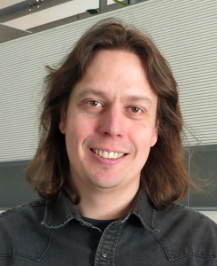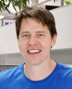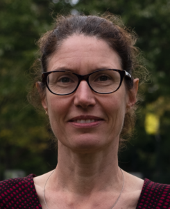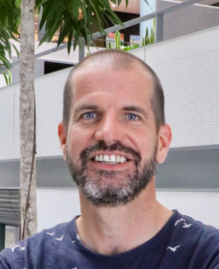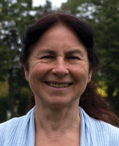
Prof. Dr. Anna Akhmanova, Utrecht University
a.akhmanova@uu.nl
Work Packages:
BioWP1.2 Cell differentiation: subcellular self-organisation and emergence of specialized cell functions
BioWP1.3 Cell migration: self-organisation during tissue rearrangement
BioWP3.1 Drug effects on tissues at the cellular and subcellular level
BioWP3.2 Interrogation and validation of drug mechanisms
TechWP1.5 Label-free sensing strategies for drugs and biomarkers
TechWP2.3 Innovative actuation strategies
Keywords:
Cytoskeleton, microtubules, membrane trafficking, in vitro reconstitution, 3D cell migration, optogenetics
Research Summary:
The Akhmanova lab studies cytoskeletal organization and trafficking processes, which contribute to cell polarization, differentiation, vertebrate development and human disease. Anna Akhmanova is an expert on microtubule dynamics and microtubule-based membrane trafficking. Current lines of research include in vitro reconstitution of the dynamics of cytoplasmic, centriolar and ciliary microtubules, microscopy-based studies of microtubule organization and function in different types of differentiated cells, analysis of microtubule contribution to different cell migration modes in 3D environments, and investigation of the mechanisms underlying the action of microtubule-targeting agents used for cancer therapy.
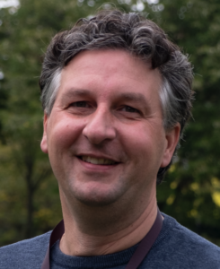
Prof. Dr. Jeffrey Beekman, University Medical Center Utrecht
j.beekman@umcutrecht.nl
Work Packages:
BioWP1.1 Cell proliferation and renewal: self-organisation
BioWP1.2 Cell differentiation: subcellular self-organisation and emergence of specialized cell functions
BioWP2.1 Normal tissue repair: de-differentiation, cell division, cell migration
Keywords:
intestinal organoids, airway organoids, biobanks, drug development, precision medicine, ion and fluid transport
Research Summary:
The research in his group focusses of the development and application of patient-derived stem cell models for basic science studies, drug development and precision medicine applications. It demonstrated that intestinal organoids from people with cystic fibrosis could guide individual treatment selection and was pivotal for developing and understanding new cystic fibrosis therapies. A current focus is to further automate and quality control adult stem cell technologies for larger-scale preclinical applications.
Dr. Federica Eduati, Eindhoven University of Technology
f.eduati@tue.nl
Work Packages:
BioWP2.3 Immunological cell clearance in regeneration and cancer
BioWP3.1 Drug effects on tissues at the cellular and subcellular level
BioWP3.2 Interrogation and validation of drug mechanisms
Keywords:
systems biology, precision oncology, mathematical modelling, microfluidics, tumour microenvironment
Research Summary:
In the Systems Biology for Oncology group we study tumours as complex ecosystems studying how diverse molecular and cellular interactions impact treatment response and tumour development holistically. Tumour response to treatment involves intricate networks within and between cells, making individual gene mutations or checkpoint molecule expression ineffective biomarkers. This challenge is even more significant for drug combinations, which help to overcome resistance. To address these issues, we investigate how tumour cells and the microenvironment (TME) influence drug response, aiming to enhance precision oncology. Our approach combines mathematical modelling and microfluidics technology.
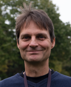
Prof. Dr. med. Peter Friedl, Radboud University Medical Center, Nijmegen
peter.friedl@radboudumc.nl
Work Packages:
BioWP1.3 Cell migration: self-organisation during tissue rearrangement
BioWP2.2 Tissue self-organisation gone awry: cancer initiation and progression
BioWP2.3 Immunological cell clearance in regeneration and cancer
BioWP3.1 Drug effects on tissues at the cellular and subcellular level
BioWP3.2 Interrogation and validation of drug mechanisms
TechWP1.2 Deeper live-imaging in mice
TechWP1.5 Label-free sensing strategies for drugs and biomarkers
Keywords:
cell migration, intravital microscopy, cancer metastasis, immune cell function
Research Summary:
His research interest is in applying innovative 3D models and engineered matrices to uncover the mechanisms and plasticity of cell migration in immune regulation and cancer metastasis, with emphasis on cell-matrix adhesion, pericellular proteolysis and cell-cell communication during migration. His laboratory identified pathways determining diversity and plasticity of cell migration, collective cancer cell invasion, and the contribution of migration pathways to immune defence and cancer resistance. His discoveries have provided a nomenclature for the different types of cell migration and their roles in building and (re)shaping tissue, with emphasis on inflammation, regeneration and cancer. His therapeutic preclinical studies focus on the intravital visualization of niches and mechanisms and strategies to overcome therapy resistance.
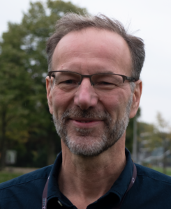
Prof. Dr. Sander van den Heuvel, Utrecht University
s.j.l.vandenheuvel@uu.nl
Work Packages:
BioWP1.1 Cell proliferation and renewal: self-organisation
TechWP1.4 Innovative labelling strategies for Expansion Microscopy
TechWP2.3 Innovative actuation strategies
TechWP2.4 Automated control of biology
Keywords:
C. elegans, cell proliferation, differentiation, spindle positioning, asymmetric cell division, (opto-)genetics
Research Summary:
Our group studies the control of cell proliferation in the context of animal development. Specifically, we focus on the process of asymmetric cell division, which supports the creation of cell diversity and maintenance of stem cells. In addition, we investigate how cell proliferation and differentiation are jointly regulated in development. An attractive model to address these questions are the mesoderm progenitors in C. elegans that go through a stereotypic pattern of cell proliferation and differentiation, to create muscle cells, scavenger cells and new progenitor cells. A multidisciplinary strategy that combines cell lineage-specific genetics, live imaging, single-cell genomics and transcriptomics, single-molecule mRNA quantifications, and theory.
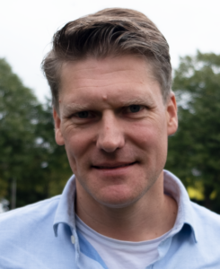
Prof. Dr. Lukas Kapitein, Utrecht University
l.kapitein@uu.nl
Work Packages:
BioWP1.2 Cell differentiation: subcellular self-organisation and emergence of specialized cell functions
BioWP1.3 Cell migration: self-organisation during tissue rearrangement
TechWP1.1 Subcellular live-imaging in transparent tissue
TechWP1.4 Innovative labelling strategies for Expansion Microscopy
TechWP2.1 Real-time data analysis
TechWP2.2 Automated control of microscopy
TechWP2.3 Innovative actuation strategies
TechWP2.4 Automated control of biology
Keywords:
cytoskeleton, super-resolution microscopy, expansion microscopy, optogenetics, feedback microscopy, epithelial polarity
Research Summary:
My group studies how cytoskeletal organisation and motor activity contribute to cellular polarisation and organelle positioning. To this end, we have developed a broad range of chemogenetic and optogenetic strategies to explore and control intracellular transport. In addition, we have pushed the boundaries of super-resolution microscopy to map the cytoskeleton of differentiated cells, such as neurons and epithelial cells.
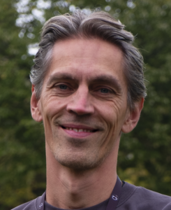
@KopsLab
Prof. Dr. Geert Kops, Hubrecht Institute
g.kops@hubrecht.eu
Work Packages:
BioWP1.1 Cell proliferation and renewal: self-organisation at the tissue level
BioWP2.1 Normal tissue repair: de-differentiation, cell division, cell migration
BioWP2.2 Tissue self-organisation gone awry: cancer initiation and progression
BioWP3.1 Drug effects on tissues at the cellular and subcellular level
BioWP3.2 Interrogation and validation of drug mechanisms
TechWP1.3 Quantitative imaging with sensors across systems
Keywords:
chromosome segregation, organoid cell biology, mitosis, cancer, advanced microscopy
Research Summary:
The main aim of the research in our lab is to understand how the cell division process gives rise to two genetically identical daughter cells. We are particularly interested in the processes that ensure correct chromosome segregation during mitosis. This is not only fascinating from a molecular perspective (how does a cell do that) and an evolutionary perspective (How has this intricate process evolved and are there meaningful differences between species?) but also has implications for health and disease. Errors in chromosome segregation are a major cause for birth defects and embryonic lethality in humans, and the most common genetic alteration in human tumors is aberrant chromosome numbers, called aneuploidy. Finally, as we and others have shown, errors in chromosome segregation during mitosis have dramatic secondary consequences on genome integrity, including translocations, deletions and chromosome shattering (chromothripsis).
Dr. ir. Arthur de Jong, Eindhoven University of Technology
a.m.de.jong@tue.nl
Work Packages:
BioWP3.1 Drug effects on tissues at the cellular and subcellular level
TechWP1.5 Label-free sensing strategies for drugs and biomarkers
Keywords:
biosensing, single-molecule, continuous monitoring, biomarkers
Research Summary:
Sensing technologies that enable the continuous monitoring of concentrations of biomolecules are in rising demand for use in dynamic biological and biomedical applications. Examples are the monitoring of patients in critical care, the monitoring of industrial processes and bioreactors, and the monitoring of ecological systems. This is what our group focuses on.
Prof. Dr. Roos Masereeuw, Utrecht University
r.masereeuw@uu.nl
Work Packages:
BioWP1.3 Cell migration: self-organisation during tissue rearrangement
BioWP2.1 Normal tissue repair: de-differentiation, cell division, cell migration
BioWP3.1 Drug effects on tissues at the cellular and subcellular level
BioWP3.2 Interrogation and validation of drug mechanisms
TechWP1.5 Label-free sensing strategies for drugs and biomarkers
Keywords:
microfluidics, organ-on-a-chip, cell differentiation
Research Summary:
My group focuses on understanding the pathways that can be pharmacologically triggered to enhance repair after organ
injury and on developing novel regenerative therapies to replace organ functions. In my lab we have developed human in vitro systems that functionally mimic (patients) organs, such as 3-dimensional advanced tissue cultures with the use of innovative technologies like microfluidics (organs-on-chip technology) and biofabrication. Target organs currently involve (but are not restricted to) the kidney and the intestine, individually and combined, allowing to study the interaction between two organs when one of them fails.
@MadelonMaurice
Prof. Dr. Madelon Maurice, University Medical Center Utrecht
M.M.Maurice@umcutrecht.nl
Work Packages:
BioWP1.1 Cell proliferation and renewal: self-organisation at the tissue level
BioWP1.2 Cell differentiation: subcellular self-organisation and emergence of specialized cell functions
BioWP2.1 Normal tissue repair: de-differentiation, cell division, cell migration
BioWP2.2 Tissue self-organisation gone awry: cancer initiation and progression
BioWP3.1 Drug effects on tissues at the cellular and subcellular level
BioWP3.2 Interrogation and validation of drug mechanisms
TechWP2.3 Innovative actuation strategies
Keywords:
Wnt signalling, organoids, cell-cell communication, cancer mechanisms, targeted membrane protein degradation
Research Summary:
The overall aim of the Maurice lab is to gain a fundamental understanding of the dual nature of signals that guide homeostatic tissue renewal and cancer cell growth. In healthy tissue renewal, a handful of signalling pathways supports the maintenance of small populations of adult stem cells. Deregulation of these pathways due to mutations is strongly linked to cancer development. Our main focus is to investigate how patient-derived mutations alter protein activity to promote the initiation and progression of cancer growth. With these insights we aim to uncover patient-specific disease mechanisms and develop improved cancer-targeting strategies. To address these issues, we combine advanced gene editing, proteomics, biochemistry and imaging approaches with organoid-based disease models.
Prof. Dr. Menno Prins, Eindhoven University of Technology
m.w.j.prins@tue.nl
Work Packages:
BioWP3.1 Drug effects on tissues at the cellular and subcellular level
TechWP1.5 Label-free sensing strategies for drugs and biomarkers
Keywords:
biosensing, quantitative imaging
Research Summary:
The Prins group aims at the development of technologies to detect proteins and study protein function with single-molecule resolution in complex macromolecular environments such as blood plasma. This entails the investigation of biophysical manipulation and detection methods with emphasis on the use of small particles. These particles offer the possibility to actively transport biological materials and to detect the presence and properties of specific targets.
The capability to probe individual molecules enables the researchers to reveal novel characteristics of the molecules, e.g. distributions of molecular properties (such as affinity or kinetic parameters, variations in space and/or in time). Moreover, it allows the study of properties that are difficult or even impossible to measure on ensembles of molecules, e.g. molecular torsion constants. This research leads to novel biosensing technologies and to a better understanding of molecular functions within complex and crowded environments.
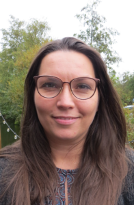
Dr. Silvia Mihăilă, Utrecht University
s.mihaila@uu.nl
Work Packages:
BioWP1.3 Cell migration: self-organisation during tissue rearrangement
BioWP2.1 Normal tissue repair: de-differentiation, cell division, cell migration
BioWP3.1 Drug effects on tissues at the cellular and subcellular level
BioWP3.2 Interrogation and validation of drug mechanisms
TechWP1.5 Label-free sensing strategies for drugs and biomarkers
Keywords:
in vitro models, biofabrication techniques, organ-on-chip, microenvironmental cues, drug testing, therapeutic targets
Research Summary:
My group specializes in creating advanced in vitro models, (kidney, gut, bone) to accurately replicate organ physiology and associated complications. Our approach involves integrating crucial components like blood vessels, immune cells, and microenvironmental factors. In our approach we combine biofabrication and bioprinting methods with cell and organoids. We apply specific microenvironmental cues, like shear stress (via perfusion) and topography (via micro and nanopatterning), to explore their impact on cellular functionality, such as baso-apical drug transporter functional polarization and barrier formation. Additionally, we assess drug efficacy, metabolite biotransformation, multi-drug interactions, and gene delivery within these models so that we uncover novel therapeutic strategies and targets. Furthermore, we use CRISPR/Cas9 to create gene knockout and knock-in models to study specific genes and create reporter cell lines and investigate their functionality when cultured under the conditions described above.
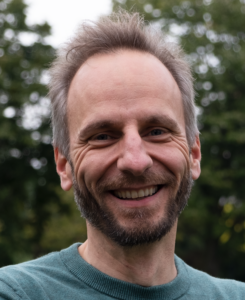
@vanRheenen_Lab
Prof. Dr. Jacco van Rheenen, Netherlands Cancer Institute, Amsterdam; Oncode institute
j.v.rheenen@nki.nl
Work Packages:
BioWP1.1 Cell proliferation and renewal: self-organisation at the tissue level
BioWP1.3 Cell migration: self-organisation during tissue rearrangement
BioWP2.1 Normal tissue repair: de-differentiation, cell division, cell migration
BioWP2.2 Tissue self-organisation gone awry: cancer initiation and progression
BioWP3.1 Drug effects on tissues at the cellular and subcellular level
BioWP3.2 Interrogation and validation of drug mechanisms
TechWP1.2 Deeper live-imaging in mice
TechWP1.3 Quantitative imaging with sensors across systems
TechWP1.5 Label-free sensing strategies for drugs and biomarkers
Keywords:
intravital microscopy, tumour initiation, treatment, metastasis
Research Summary:
The van Rheenen group studies the identity, behavior, and fate of cells that drive tumour initiation, progression, metastasis and the development of therapy resistance. These populations of cells are difficult to study since they are rare, and their behaviour (e.g. migration) and traits (e.g. stemness) change over time. To be able to study these dangerous cells, we have developed intravital microscopy. Using intravital imaging, the van Rheenen lab revealed multiple important factors within the single cell heterogeneity that are crucial in the processes of tissue homeostasis, tumour initiation and tumour progression.
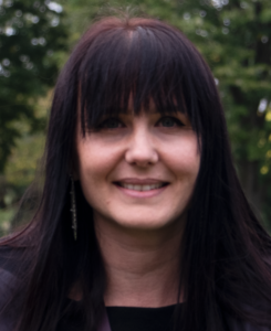
@Dream3DLab
Prof. Dr. Anne Rios, Princess Máxima Centre of Pediatric oncology, Utrecht
A.C.Rios@prinsesmaximacentrum.nl
Work Packages:
BioWP2.2 Tissue self-organisation gone awry: cancer initiation and progression
BioWP2.3 Immunological cell clearance in regeneration and cancer
TechWP2.1 Real-time data analysis
TechWP2.2 Automated control of microscopy
TechWP2.4 Automated control of biology
Keywords:
multispectral 3D imaging, organoids, computer vision; human biology; cancer immunotherapy
Research Summary:
The Rios Dream3dLab is a collaborative team that develops technology in the field of in vitro human modelling, 3D imaging, and computer vision. Using these advanced tools, they create a patient-centric immuno-oncology platform, allowing for a comprehensive analysis of the effectiveness and varying responses to cancer immunotherapy. Their overarching objective is to develop customized, next-generation cancer treatments that tailor to specific patient populations. Beyond science, they are committed to making a positive impact on society. They promote sustainability through the Green Lab, support diversity via community-building initiatives like ACE, which empowers women in science. They are also passionate about engaging the public in science through immersive microscopy experiences.
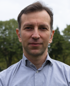
Dr. Ihor Smal, Utrecht University
i.v.smal@uu.nl
Work Packages:
TechWP1.2 Deeper live-imaging in mice
TechWP2.2 Automated control of microscopy
TechWP2.4 Automated control of biology
Keywords:
smart microscopy, image analysis, tracking and explainable AI
Research Summary:
Our group focuses on development of novel approaches for real-time image processing and data analysis using statistical signal processing and modern AI approaches to enable automated real-time manipulation of key aspects of the biological systems. Those solutions will be used for automated real-time optimization of imaging experiments, to control cellular architecture, migration and fate with light and provide feedback to the imaging systems to achieve automated guidance of biological processes.
Dr. Carlas Smith, Delft University of Technology
c.s.smith@tudelft.nl
Work Packages:
TechWP1.1 Subcellular live-imaging in transparent tissue
TechWP1.2 Deeper live-imaging in mice
TechWP2.2 Automated control of microscopy
TechWP2.4 Automated control of biology
Keywords:
computational imaging, smart microscopy, super resolution, quantitative imaging, image analysis
Research Summary:
Our research group is dedicated to pushing the boundaries of nanoscopy resolution to a point where it becomes feasible to extract vital information about biomolecular actions and interactions within intact live tissue. We firmly believe that the time is ripe, both in terms of technological advancements and conceptual innovations, to embark on the journey from achieving optical imaging at unprecedented resolutions to establishing ‘functional nanoscopy of biomolecular processes.’ Over the upcoming years, we will collaborate closely with scientists who employ these cutting-edge technologies, working in tandem to advance optical nanoscopy technology. Together with our esteemed collaborators, our mission is to explore the living brain, with the ultimate goal of uncovering the intricacies of memory formation and the processes underlying learning.
Dr. Hugo Snippert, Princess Máxima Centre of Pediatric oncology, Utrecht; Oncode Institute
h.j.g.snippert-2@prinsesmaximacentrum.nl
Work Packages:
BioWP2.1 Normal tissue repair: de-differentiation, cell division, cell migration
BioWP3.1 Drug effects on tissues at the cellular and subcellular level
BioWP3.2 Interrogation and validation of drug mechanisms
TechWP1.3 Quantitative imaging with sensors across systems
TechWP2.1 Real-time data analysis
Keywords:
human organoids, biosensors, molecular genetics, live-cell microscopy, genomics
Research Summary:
Cells within a cancer are highly heterogeneous with respect to their phenotype and can manifest distinct morphological, molecular and functional features. As a consequence, it is challenging to design treatment therapies that target all cancer cells as effectively.
Using human samples of colorectal cancers, the Snippert lab studies the causes and consequences of heterogeneity in cellular behaviour during tumour growth, tumour evolution and the emergence of therapy resistance. Primarily, we study tumour cells using cancer organoids and live-cell imaging experiments to assess changing cell states, real-time signalling dynamics and evolving genomes, all with single-cell resolution.
Prof. Dr. Annemiek van Spriel, Radboud University Medical Center, Nijmegen
annemiek.vanspriel@radboudumc.nl
Work Packages:
BioWP1.2 Cell differentiation: subcellular self-organisation and emergence of specialized cell functions
BioWP2.3 Immunological cell clearance in regeneration and cancer
BioWP3.1 Drug effects on tissues at the cellular and subcellular level
BioWP3.2 Interrogation and validation of drug mechanisms
Keywords:
immunology, membrane proteins, tetraspanins, signal transduction, super-resolution imaging, tumour models
Research Summary:
The plasma membrane has a unique spatial organization of proteins and lipids that is critical for cell structure and function. Tetraspanin proteins, expressed in the plasma membrane of virtually all mammalian cells, coordinate the spatial organization of specific proteins and signalling molecules into ‘tetraspanin nanodomains’. We discovered that tetraspanins control fundamental cellular processes in immune cells, including proliferation and antigen presentation. The aim of our group is to unravel the molecular mechanisms that underlie tetraspanin nanodomain function in immune cells in relation to the development of malignant diseases.
Prof. Dr. Sjoerd Stallinga, Delft University of Technology
s.stallinga@tudelft.nl
Work Packages:
TechWP1.1 Subcellular live-imaging in transparent tissue
TechWP1.2 Deeper live-imaging in mice
TechWP2.2 Automated control of microscopy
Keywords:
super-resolution microscopy, computational imaging, optical design
Research Summary:
My research focuses on the intersection between the fundamentals and engineering of optical imaging systems and image processing algorithms (“computational imaging”). This combination makes it possible to see what cannot be made visible with conventional optical imaging instrumentation. We apply this to super-resolution microscopy to achieve nanometre resolution, and to develop automated slide scanning solutions for digital pathology.
Dr. Martijn Verdoes, Leiden University Medical Center, Leiden
m.verdoes1@lumc.nl
Work Packages:
BioWP2.2 Tissue self-organisation gone awry: cancer initiation and progression
BioWP2.3 Immunological cell clearance in regeneration and cancer
TechWP1.4 Innovative labelling strategies for Expansion Microscopy
TechWP2.3 Innovative actuation strategies
Keywords:
chemical immunology, targeted immunomodulation, immunotherapy, activity-based probe design
Research Summary:
The Verdoes lab conducts multidisciplinary science at the interface of chemistry and immunology to gain new insights for the development of more effective and safer immunotherapeutic approaches.
