This gallery features selected (microscope) images and videos acquired by our PhDs and postdocs.
Microscope: Nikon spinning disk confocal microscope.
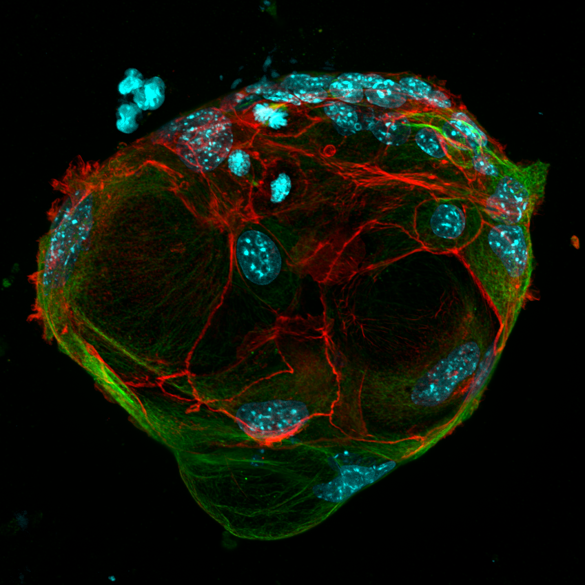
– Alessandro Corsini
(Blue = DAPI, Red = Phalloidin, Green = P-gp)
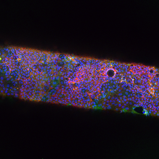
– Ralph de Groot
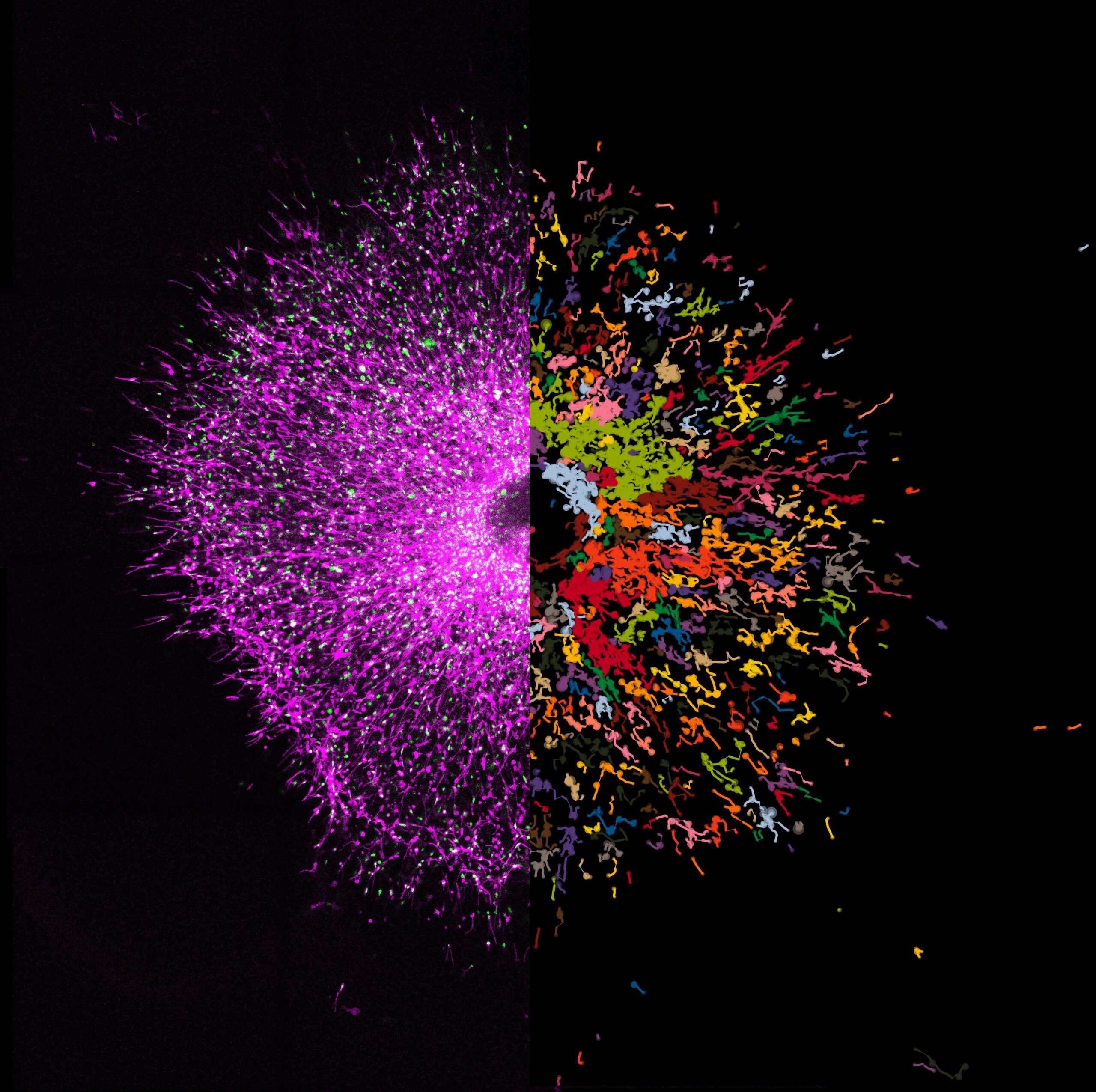
– Stijn den Daas
(Cyan = DAPI, Magenta = Cleaved-caspase-3)
– Tatum van Maanen
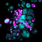
Microscope: Nikon spinning disk confocal microscope.
– Alessandro Corsini